
Umbilical Cord Wrapped Around Neck
The term “nuchal chord” refers to a condition when umbilical cord wrapped around neck of baby while still in the womb. Let’s discuss about the umbilical cord and various abnormalities.
The finish or umbilical cord forms the connecting link between the fetus and placenta. It is through this umbilical cord that the fetal blood flows to and from the placenta. It extends from the fetal umbilicus to the fetal surface of placenta.
Development of Umbilical Cord
It is developed from body stalk of mesoderm tissue stretching between embryonic disc and chorion.
Characteristicsof Umbilical Cord
(a) It is 50 cm in length with a variation of 30-100 cm. It is bluish white in colour.
(b) Its diameter varies from 1-2.5cm. It is twisted spirally from left to right-40 twists.
(c) Its thickness is not uniform but presents notes or swelling at places. These swellings are due to dilation of umbilical veins. Or local collection of Wharton’s jelly. These swellings are also called false knots.
(d) Initially two arteries and two veins form. Arteries have connections with fetal internal illiac arteries. They carry venous blood from fetus to placenta. Of two umbilical veins developed, right one appears by 16th week thus single vein remains carrying oxygenated blood from placenta to fetus.
(e) The Umbilical arteries do not possess internal elastic lamina but have got well developed muscular coat. This helps in effective closure of arteries due to reflex spasm soon after the birth of baby.
Structures of Umbilical Cord
The structures of Umbilical Cord are listed below:
- Covering epithelum
- Wharton’s jelly
- Blood vessels
- Remnant of Umbilical vesicle (Yolk sac) and its vitelline duct.
- Allantois.
- Obliterated extra embryonic coelom.
- Covering Epithelium: Lined by single layer of amniotic epithelium. But at term shows stratification like that of fetal epidermis at birth.
- Wharton’s Jelly: It is rich Mucopolysaccharides and has got protective function to the umbilical vessels.
- Blood Vessels
(a) Initially there are 4 vessels, 2 arteries and 2 veins.
(b) The arteries are derived from the internal illiac arteries of the fetus and carry the venous blood from the fetus to placenta.
(c) Of the 2 Umbilical veins, the right one disappears by the 4th month, leaving behind one vein which carries oxygenated blood from placenta to fetus. Presence of a single umbilical artery is often associated with fetal congenital abnormalities.
- Remnant of the Umbilical Vesicle (Yolk sac) and its Vitelline Duct: It is found as small yellow body near the attachment of the cord to the placenta or on rare occasion, the proximal part of the duct presists as Meckel’s Diverticulum.
- Allantois: A blind tubular structure may ocassionally be present near the fetal end which is continuous inside the fetus with its Urachus and bladder.
- Obliterated Extra Embryonic Coelom: Intra- embryonic coelom is continuous with extra embryonic coelom along with herniation of coil of intestine (midgut). The condition may persist as congenital Umbilical hernia or Examphlous.
Attachment
Umbilical cord is centrally attached on fetal surface of placenta and at the umbilicus of fetus.
Functions of Umbilical Cord
- Lifeline between placenta and fetus supplying oxygen and nutrients to fetus and disposing waste products.
- Exchange of fluid and electrolyte between umbilical vessels and the amniotic fluid.
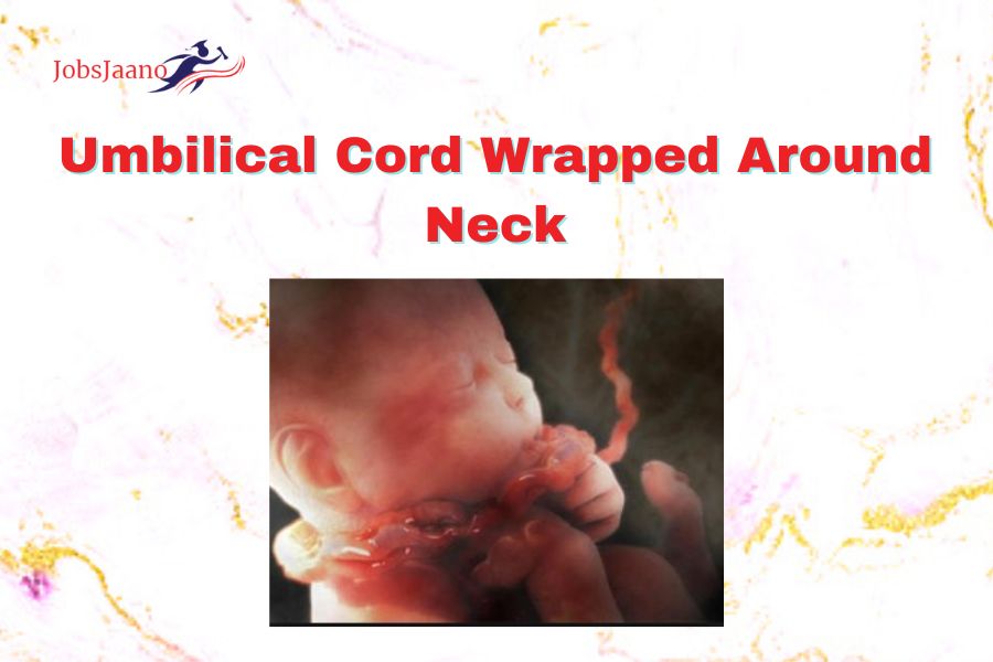
Umbilical Cord Around Neck
Your midwife or obstetrician will likely cut the umbilical cord (nuchal cord) if it becomes wrapped around your baby’s neck before you even realise it. It happens in up to a third of births, which is shockingly common, and it’s quite unlikely that it will harm you or your kid.
During birth, a nuchal chord frequently goes totally undiscovered and unaffected. However, your midwife can quickly correct it if she discovers the umbilical cord around your baby’s neck after his head has been delivered. Either she will slip the cord over your baby’s head or she will simply loosen the cord to allow your baby’s shoulders to pass through.
Your midwife may clamp and cut the cord if it is tightly wrapped around your baby’s neck before his shoulders are born. However, it’s uncommon for this to be required.
Your baby’s heart rate will allow your midwife to determine if there are any problems with blood flow in the cord. The umbilical cord can occasionally become crushed during a contraction, which may cause a transient decrease in his heart rate.
Your midwife might ask for permission to closely monitor your baby’s heartbeat if she has reason to believe that this is taking place. Your labour can continue unabated as long as he is being watched and there are no more issues.
Try to remain calm. The midwife should explain everything to you and your spouse because issues like these are uncommon.
Speak with your midwife if you have any worries; she will be able to reassure you.
Abnormalities of the Umbilical cord
The abnormalities of the cord are:
(a) Short Cord
(b) Long Cord
(c) True Knot
(d) False Knot
(e) Loops of Cord
(f) True Cyst of Cord
(g) False Cyst.
(a) Short Cord: It is less than 30cm (12 inches)
(i) There can be no cord (achordia)
(ii) Breech presentation favours shorter cord.
(iii) It causes abruption Placenta & uterine inversion.
(b) Long Cord: It is more than 100cm (40 inches)
(i) It can be as long as 300cm.
(ii) Long cord develops true knot and prolapse cord.
(c) True Knot: Occurs in 1% & is rare.
(i) Common in a monoamniotic twins.
(ii) Perinatal loss increases three fold.
(d) False Knot: Occurs from kinking of vessels and a developmental variation.
(i) It has no clinical importance.
(ii) It occurs due to an accumulation of whartons jelly.
(e) Loops of umbilical cord: They are of 3 types:
(i) One loop of cord around fetal neck (21%)
(ii) Two loops of cord around fetal neck (3.5%)
(iii) Three loops of cord around fetal neck (0.2%)
(f) True cyst of cord: Occurs due to remnant of umbilical vesicle or of allntois.
(g) False Cyst: Liquification of wharton’s jelly.
Single Umbilical Artery: It is present in about 1% cases more common in twins & babies born to diabetic mother. It is often associated with congenital malformation of the fetus. There are increased chances of abortion prematurly, dysmaturly & increased perinatal mortality.