
Diagnosis Of Pregnancy From 1-12 Weeks (First Trimester)
Pregnancy Diagnosis Test
It can be diagnosed by the (A) Subjective and (B) Objective Symptoms,
(A) Subjective Symptoms
These all symptoms appear early in the pregnancy.
they include:
- Amenorrhea.
- Morning Sickness.
- Frequency of Micturition.
- Breast discomfort.
- Fatigue.
(B) Objective Signs
These include:
- Breast Changes.
- Changes per abdomen that is the pelvic Changes.
- Immunological tests.
(A) SUBJECTIVE SYMPTOMS
-
AMENORRHEA symptoms, causes, treatment:
It means missing of menses.
(a) Cyclic bleeding may occur upto 12 weeks of pregnancy until the decidual space is obliterated by the fusion of decidua vera with decidua Capsularis.
(b) Such bleeding is usually scanty, Lasting for a shorter duration and is not to be confused as last menstruation in calculating duration of pregnancy. This is also called Placental sign or implantation bleeding.
- MORNING SICKNESS
It is present in 50% cases more in 1st pregnancy. It usually appears soon after the missed period and rarely lasts beyond 3rd month.
(a) It varies from Nausea on rising from the bed to loss of appetite or even vomiting.
(b) But it does not affect the health status of the mother.
(c) It passes off after 100 days.
- FREQUENCY OF MICTURATION
(a) It appears during 8-12th week of pregnancy.
(b) It is quite troublesome.
(c) As the uterus straightens up after 12th week symptoms disappear.
- BREAST DISCOMFORT
It is evident as early as 6-8 weeks with feeling fullness and pricking sensation.
- FATIGUE
It occurs early in pregnancy.
BREAST CHANGES
(a) These are more clear in primigravida. (b) Breast changes occur between 6-8 weeks.
(c) There is breast enlargement with vascular engorgement and due to this delicate veins and visible under the skin.
(d) The nipple and areola (Primary) become pigmented.
(e) Montgomery tubercle’s are prominent.
(f) And above all from the 12th week thick yellowish secretion (Colostrum) can be expressed.
CHANGES PER ABDOMEN THAT IS PELVIC CHANGES
Uterus remains a pelvic organ until 12th week, reafter it is felt per abdomen as a suprapubic bulge.
PELVIC CHANGES
These changes are diverse and appear at different iods. These are:
Jacquemier’s or Chadwick sign. Vaginal sign including (Osiander’s sign) Cervical signs (including Goodell’s sign) Uterine Signs (Piskacek’s sign in pregnancy)
Hegar sign.
Palmar sign.
These all signs are explained one by one:
Jacquemier’s Or Chadwick Sign:
Weeks of Appearance of chadwick sign: 8th week of Pregnancy.
What Happens: It is the dusky hue of vestibule and anterior vaginal wall.
Appears more in multiparae.
Causes: It is due to local vascular congestion and may be brought about by pelvic tumor i.e uterine fibroid.
Vaginal Sign including Osiander Sign: (Osiander sign in pregnancy)
(i) Weeks of appearance: 6th week-8th week onwards.
(ii) What happens: Apart from bluish discolouration of anterior vaginal wall, the walls become soft and copious non-irritating mucoid discharge appears at 6th week. This is known as vaginal sign. Similarly At 8th week there is increased pulsation felt through the lateral fancies called Osiander Sign.
Note: One should not get confused with the Osiander Sign as same pulsation is felt in Acute Pelvic Inflammation.
Cervical Sign or Goodell’s Sign:
(i) Appearance of cervical/ Goodell’s Sign: 6th week.
(ii) What happens: Cervix becomes soft as early as 6th week (Goodell’s Sign). Around the exterior OS and in the upper portion, there is a noticeable softness.
Uterine Signs:
Including (Piskacek’s Sign)
(i) Size: The uterus is enlarged to the size of hen’s egg at 6th week.
Cricket ball at 8th week.
& Size of Fetal head by 12th week.
(ii) Shape: The pyriform shape of non-pregnant uterus becomes globular by 12 weeks. At 6-8 wks it becomes acutely anteverted.
(iii) Sign: Piskacek’s Sign: If there is lateral implantation there may be asymmetrical enlargement of uterus. This is referred to as the Piskacek sign, where one part is firmer than the other.
(e) Hegar Sign: Hegar Sign is demonstrated between 6-10 weeks. Its foundation is the following:
(i) Upper part of the body of uterus is enlarged by the growing ovum.
(ii) Lower part is empty and extremely soft.
(iii) & Cervix is comparatively firm.
(f) Palmer’s Sign: It is written below: Regular and rhythmic uterine contraction can be elicited during bimanual examination as early as 4-8 weeks.
-
IMMUNOLOGICAL TESTS | PREGNANCY TESTS
This is done by
- Urinary Immunological Test.
- Ultrasonography.
- Urinary Immunological Test
(a) Latex Agglutination Slide test.
(b) Immunochromatographic test.
(c) Latex Agglutination Slide Test.
Inhibition of agglutination in the slide test indicates a positive result.
(b) Immunochromatographic Test
It is more sensitive than former test at 20-50 U/ml after 1 week of missed period.
ELISA or Radio immunoassay (RIA)-It is doing for diagnosis of early pregnancy. Its special indication is in follow-up of patients with trophoblastic disease. This test is done mainly for detection of Beta hC in serum/urine upto 5mlU/ml.
It can detect Beta hC G on 8th day of fertilization before periods are missed.
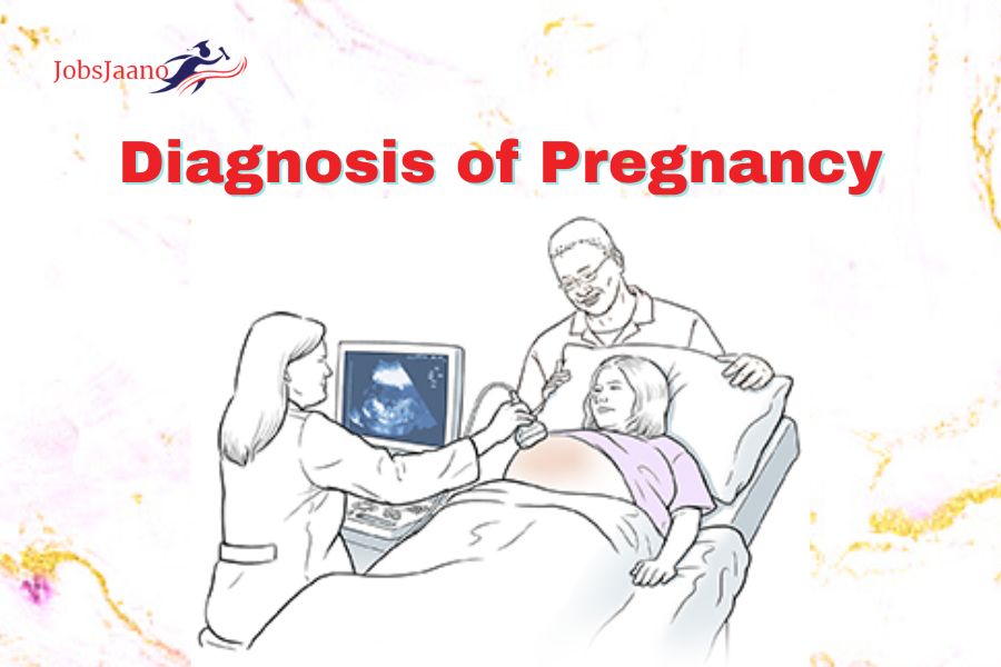
- Ultrasonography
(a) Abdominal USG can detect pregnancy at earliest e.
(b) Spherical Gestation Sac-5th week.
(c) Fetal pole- 6th week.
(d) Crown-Rump length 7th week.
(e) Visualisation of fetal heart motion-7th week.
(f) Biparietal diameter (BPD) 2.1 cm at 12th week.
(g) Fetal heart Sound 10th week by ultrasound doppler.
(h) Transvaginal Sonography (TVS)
Can diagnose earlier than abdominal sonography
as
Gestational Sac – 4th Week.
Yolk Sac – 5th Week.
Cardiac Motion – 5th Week.
Fetal Pole – 5th Week.
SECOND TRIMESTER OR 4-7 MONTHS
can diagnose pregnancy of 13-28 weeks as follows:
Symptoms.
General Examination.
Abdominal Examination.
Vaginal Examination.
Investigation.
SYMPTOMS
- Amenorrhea Continues.
- Quickening is present. It is also known as feeling of life. Quickening denotes the perception of active fetal movements by the women.
- It is felt at 18th week & useful guide to calculate the expected date of delivery.
Note: Quickening occurs at 18th week in Primigravida & at 16th week in Multigravida. In the end there is progressive enlargement of lower abdomen from 5th month.
GENERAL EXAMINATION
On General Examination we see the following:
Chloasma
It appears at about 24th week. Chloasma is the pigmentation over the forehead, face and Cheeks.
Breast Changes
Breasts are larger and under the skin there are more noticeable veins.
Secondary Areola appears at about 20th week.
By the sixteenth week, the colostrum is thick and yellowish.
The conspicuous Montgomery tubercles reach the secondary areola.
Abdominal Examination
It includes the following:
(i) Inspection
Linea nigra appears at 20th week. It extends from symphysis pubis to ensiform cartilage. Striae (Both pink & white) of varying degree become visible in the lower abdomen, move towards the flanks,
(ii) Palpation
Fundal height is increased with the enlargement of uterus.
The following formula is a useful guide for calculating the fundal height.
At 16th week: The height of uterus is midway between symphysis pubis and Umbilicus.
At 24th Week: It is at the level of Umbilicus.
At 28th Week: It is present at the junction of lower third and upper two-third of distance between the Umbilicus and ensiform cartilage.
(a) In second trimester the uterus feels soft, elastic and becomes ovoid in shape.
(b) Braxton Hicks Contractions too appear. (c) By 20th week fetal parts can be palpated, hence it helps in noting the presentation & position of fetus in later weeks.
(d) At early 20 weeks active fetal movements are felt.
(e) External ballotment is elicited as early as 20th week.
AUSCULTATION
(a) Fetal heart sound is the most conclusive clinical sign of pregnancy. It is detected between 18-20 weeks.
(b) The rate of F.H.S varies from 140-160 per minute but gradually settles down to 120-140 per minute as the pregnancy advances.
VAGINAL EXAMINATION
(a) On Vaginal Examination there is bluish discolouration of Vulva, Vaginal. Cervix & softening of cervix.
(b) Between 16-28th weeks Internal balloment can be elicited.
INVESTIGATIONS
Mainly 2 Investigations are done in the 2nd trimester.
(a) Sonography: Routine Sonography is done at 18- 20 weeks for detailed fetal anatomy, Placental site and integrity of cervical canal.
(b) Radiography: It is done at early 16th week & in this the fetal skeletal shadow is visible.
LAST TRIMESTER (29-40 WEEKS)
It can be diagnosed by the following
SIGNS & SYMPTOMS
Signs
(a) Cutaneous changes appear with increased pigmentation and striae.
(b) Uterine shape is changed from cylindrical to spherical after 36 weeks.
(c) Fundal height increases further as follows:
(i) At 32 weeks: The fundal height corresponds to the junction of upper and middle third at 32 weeks.
(ii) At 36th Week: Upto the level of Ensiform cartilage at 36 weeks.
(iii) 40 Weeks: It comes down to the level of 32 weeks due to engagement of head.
Note: If the head is floating it is of 32 weeks of pregnancy and if head is engaged it is of 40th week pregnancy.
(d) Braxton Hicks Contractions are more evident
(e) Fetal Movements are easily felt.
(f) Palpation of fetal parts is much easier and in helps in the determination of Lie, presentation and presenting part.
(g) F.H.S. is heard.
(h) Sonography.
Symptoms
The following symptoms appear in the last trimester.
(a) Amenorrhea continues.
(b) There is progressive enlargement of abdomen which causes mechanical discomfort to the pregnant woman i.e palpitation or dyspnoea following exertion.
(c) Lightening occurs at about 38th week special in primigravida. Lightening is a sense of relief of the Pressure symptoms due to the engagement of the presenting part.
(d) Frequency of Micturation re-appears.
(e) Fetal movements are more pronounced.
FAQs
Q.1. When does the Reproductive period of a woman begin?
Q.2. Write the duration of pregnancy in months, days and weeks?
Q.3. How is the duration of pregnancy calculated?
Q.4. Define Gestational age or Menstrual age?
Q.5. How is the True Gestational Age Calculated?
Q.6. For whom is the fertilization or Ovulatory age important?
Q.7. What is the duration or time period of first trimester?
Q.8. How is first trimester diagnosed? It is diagnosed by the following:
Q.9. What are the subjective symptoms?
Q.10. Of what clinical condition is the placental sign Suggestive?
Q.11. What is placental sign?
Q.12. When does frequency of Micturation appear?
Q.13.Why does frequency of micturation occur?
Q.14. When do the symptoms of frequency of Micturition disappear?
Q.15. What all changes appear in objective signs?
Q.16. What all appear in the accessory organs (Breasts)?
Q.17. Define Chadwick/Jacquemier sign?
Q.18. Why does it appear?
Q.19. With what can Osiander sign be confused?
Q.20. What is Hegar Sign?
Q.21. How is it done?
Q.22. What care should be taken while eliciting Hegar sign?
Q.23. Define Quickening or feeling of life.
Q.24. What is Chloasma or (Pregnancy mask)
Q.25. When does colostrum become thick & yellowish ?
Q.26. What is Lightening and when does it appear?
Q.27. How will you differentiate between the fundal height of 32 weeks and 40 weeks?
Q.28. How will you predict the gestational age with greater accuracy?
Q.29. How will you calculate the weight of fetus using Johnson's Formula?
Related Queries: